
Poster Download: Detection of Antibody-Secreting Cells in Cyto-Mine® Chroma
Comparison of Bead-Based and FRET Antigen-Specific Assays
Download the Poster NowPoster Overview
Efficient identification of antigen‑specific antibody‑secreting cells (ASC) is critical for antibody discovery. Cyto‑Mine® Chroma integrates picodroplet technology, multi‑laser detection, and single‑cell analysis to enable high‑throughput screening and isolation of secreting cells. In this poster, we compare three antigen‑specific assay formats-two bead‑based approaches and a FRET assay -and show they deliver comparable sensitivity and accurate detection of antigen‑specific ASC.
What you’ll learn
- How Cyto‑Mine® Chroma encapsulates, incubates, screens, sorts, and dispenses viable single cells in picodroplets.
- How FRET and bead‑based assays are configured for antigen‑specific ASC detection (including gating considerations and background controls).
- Performance comparison across assays for rare-cell detection (1:100 spike‑in) and strategies to reduce non‑specific binding.
Key Findings (at-a-glance)
- Cyto‑Mine® Chroma enables multiplexed picodroplet analysis with 4 lasers and 4 detectors for flexible assay design.
- Antigen‑specific FRET assay detected antigen‑specific ASC at a frequency consistent with IgG secretion detection in the same system (2‑hour incubation).
- Both bead‑based formats sensitively detected rare TNF‑α‑specific ASC in a 1:100 spike‑in and performed comparably to antigen‑FRET under matched conditions.
- Blocking Fc and B-cell receptor interactions, and excluding non-viable cells reduced non‑specific binding and maintained low background in controls.
Assays
We evaluated three antigen‑specific assays for detecting TNF‑α‑specific ASC on Cyto‑Mine® Chroma: (1) an antigen‑specific FRET assay using labelled antigen and anti‑IgG probes; (2) an anti‑mouse IgG (Fc) capture bead format with fluorescent antigen readout; and (3) a streptavidin bead format loaded with biotinylated antigen and fluorescent anti‑IgG detection. Across formats, antigen‑specific events were detected with comparable sensitivity, supporting flexible assay selection depending on workflow needs.
Assay Formats Compared
Bead‑based assay (anti‑mouse IgG (Fc) capture beads)
Anti‑mouse IgG‑coated beads capture secreted IgG; fluorescent TNF‑α binds captured antibody for localized bead signal.
Readout: localized fluorescent signal on beads within droplets; FcR blocking, viability gating, and sample preparation strategy reduces false positives.
Bead‑based assay (streptavidin beads + biotinylated antigen)
Streptavidin beads loaded with biotinylated TNF‑α capture antigen‑specific antibodies; fluorescent anti‑IgG detects bound antibody.
Readout: localized fluorescent bead signal; FcR + BCR epitope blocking, viability gating, and sample preparation strategy minimized false positives
Antigen‑specific FRET assay
Labelled TNF‑α antigen (donor) + labelled anti‑mouse IgG (acceptor) generate a FRET signal when both bind secreted antigen‑specific IgG.
Readout: FRET‑positive picodroplets during sorting; enable direct antigen specificity detection.
The Workflow:
Step 1
Co-encapsulate cells + assay reagents into picodroplets.
Step 2
Incubate to allow antibody secretion and signal accumulation (2 hours in this study).
Step 3
Encapsulate single cells in picodroplets with optimized probe ratio
Step 4
Screen picodroplets with multi‑laser detection to identify antigen‑specific events
Step 5
Dispense antigen-specific ASC for downstream analysis (e.g. RT-PCR)
Conclusions
Two bead-based assays were developed to detect TNF‑α‑specific ASC on Cyto‑Mine® Chroma: (i) anti‑mouse IgG (Fc) capture beads and (ii) streptavidin beads loaded with biotinylated TNF‑α.
Both bead formats enabled sensitive detection of antigen‑specific ASC in picodroplets and maintained low background with appropriate gating, blocking, and sample preparation strategy.
Under matched conditions, bead‑based assays performed comparably to the antigen‑FRET assay for detection of rare antigen‑specific ASC.
Together, the results highlight Cyto‑Mine® Chroma as a robust, flexible platform for antigen‑specific ASC screening within antibody discovery workflows.
Want the full poster?
Title: Detection of Antibody-Secreting Cells in Cyto-Mine® Chroma: Comparison of Bead-Based and FRET Antigen-Specific Assays
Authors: Maryam Ahmadi, Jitender Bisht, Elena Shvets, Eric Jabart, Caroline Falzone and Richard Hammond

Keywords: Cyto‑Mine® Chroma; picodroplet technology; microfluidics; antibody discovery; antibody‑secreting cells; antigen‑specific screening; FRET; bead‑based assay; single‑cell analysis; high‑throughput screening.
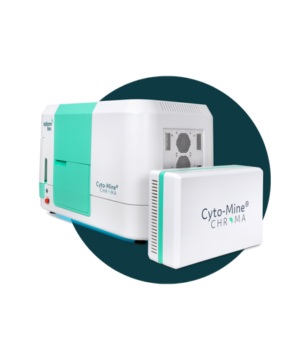
The Next Generation: Cyto-Mine® Chroma
Automate. Accelerate. Analyze millions of cells in a single day.
Think Cyto-Mine®, but supercharged, enabling multiplexing and greater assay flexibility to fit your needs. It means that you can examine vastly greater numbers of cells — and isolate the most valuable ones— with unparalleled precision.