

Protocol: Antigen Specific anti-mouse IgG (Fc) Bead-Based Assay For Use With Cyto-Mine® Chroma
Download Now
Protocol Overview
Designed for use with our Cyto‑Mine® Chroma platform, this protocol supports confident identification of rare, high-value antibody candidates while streamlining antibody discovery workflows.
By following this protocol developed by our in-house experts, you can expect to achieve high-throughput, efficient detection and isolation of rare antigen-specific antibody-secreting cells (ASCs) within heterogeneous populations, including rare populations present at low frequency (~1%).
Learn how to create an Fc bead-based assay together with approaches designed to reduce false positive signal and background noise. This assay uses anti-mouse IgG (Fc)-coated beads to capture secreted antibodies, followed by detection using fluorescently labelled antigen.
We’ve also combined the use of a viability probe with the bead-based detection so that apoptotic or compromised cells can be excluded. This is particularly important in bead-based assays, where non-specific binding from non-viable cells can significantly increase background signal and false positives.
We used well characterized control experiments, including secretion detection and antigen-specific binding using FRET assays to verify the sensitivity and accuracy of the technique.
Direct comparison shows that the Fc bead-based assay delivers performance comparable to antigen-specific FRET, with similar sensitivity and specificity for identifying rare secreting cells.
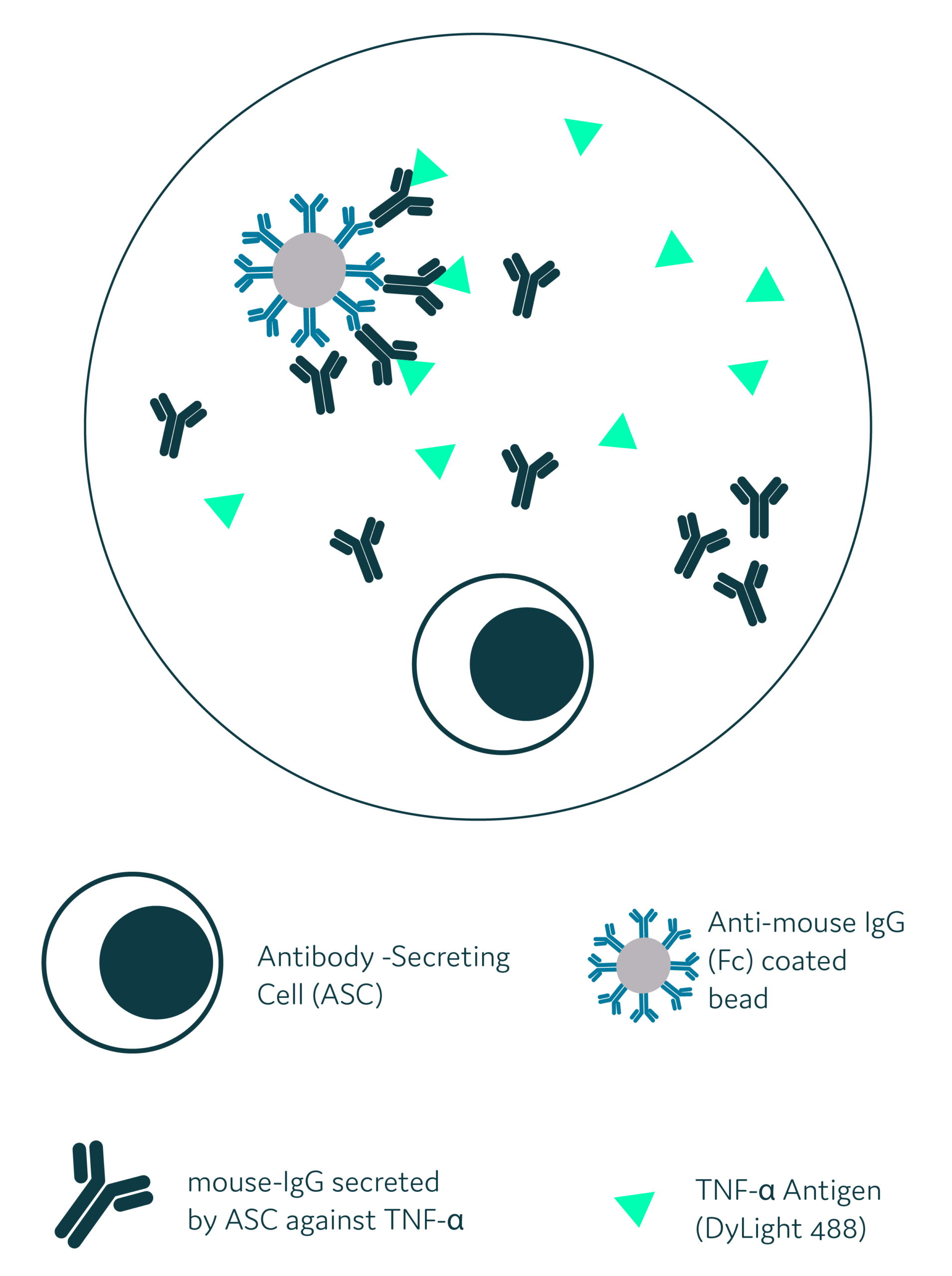
Schematic diagram for Antigen-Specific anti-mouse (Fc) Bead-Based Assay: Antibody-secreting cells (ASC) are co-encapsulated with anti-mouse IgG (Fc)-coated beads and fluorescently labelled antigen (e.g. TNF-α). Upon secretion antibodies bind to the Fc-coated bead surface and the captured antibody subsequently interacts with the fluorescent antigen. This results in the formation of a localized fluorescent complex on the bead surface within the picodroplet.
Have you read enough to want the full protocol?
If yes, simply click the button, and fill in the form to download the complete protocol and start implementing antigen-specific bead-based screening on Cyto-Mine® Chroma.
Otherwise, keep reading if you need a little more information about the workflow and some of the key benefits first!
How it Works:

Step 1
Prepare viability probe and bead-based assay
Step 2
Single‑Cell Encapsulation:
- ASCs are encapsulated into uniform picodroplets with Fc beads and fluorescent antigen
Step 3
Incubation & Signal Generation:
- Secreted antibodies are captured on Fc-coated beads and bind fluorescent antigen, generating a measurable localized emission signal
Step 4
Gating & Sorting:
- Gate 1: Exclude non‑viable cells
- Gate 2: Include IgG‑secreting viable cells
Step 5
Dispense antigen-specific ASC for downstream analysis (e.g. RT-PCR)
Key Benefits:
1) Early Identification of Rare Clones
Capture low-frequency antigen-specific ASCs early in the discovery pipeline
2) High Throughput detection of Antigen-specific clones
Screen millions of cells in a single run, reducing development timelines and costs
3) Reduced false positive / background noise
Step-by-step instructions for sample preparation to reduce false positive responses during detection and isolation of clones using Cyto-Mine® Chroma
4) High Specificity
Clear, localized fluorescence signals support accurate identification of antigen-specific events
5) Flexible & Adaptable
Develop and optimize multiple assay formats on Cyto-Mine® Chroma to suit your application
Ready for the full protocol now?
Simply click the button, and fill in the form to download the complete protocol and start implementing antigen-specific bead-based screening on Cyto-Mine® Chroma.
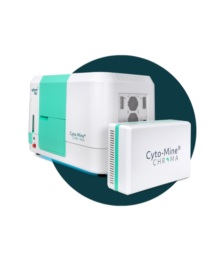
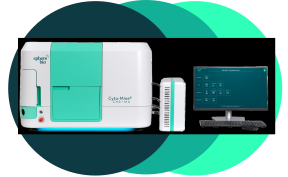
The Next Generation: Cyto-Mine® Chroma
Automate. Accelerate. Analyze millions of cells in a single day.
Think Cyto-Mine®, but supercharged, enabling multiplexing and greater assay flexibility to fit your needs. It means that you can examine vastly greater numbers of cells — and isolate the most valuable ones— with unparalleled precision.