

Protocol: Use of Cyto-Cellect® Fc Fusion and Viability Probe with Cyto-Mine® Chroma
Download Now
Protocol Overview
Authors: Elena Shvets, Jitender Bisht, Maryam Ahmadi, Claire Cox, and Richard Hammond.
Using the Cyto‑Cellect® Mouse assay kit in conjunction with our Viability Probe provides a powerful, multiplexed FRET‑based method enabling simultaneous detection of mouse IgG secretion and cell viability at the single‑cell level on the Cyto‑Mine® Chroma platform.
It delivers high‑throughput, automated single‑cell screening on the Cyto‑Mine® Chroma platform, improves hit quality by removing compromised cells early, and provides sensitive, specific secretion detection through donor‑acceptor FRET pairing – all of which streamline discovery, enhance downstream sequencing and expansion success, and accelerate the development of strong candidate clones.
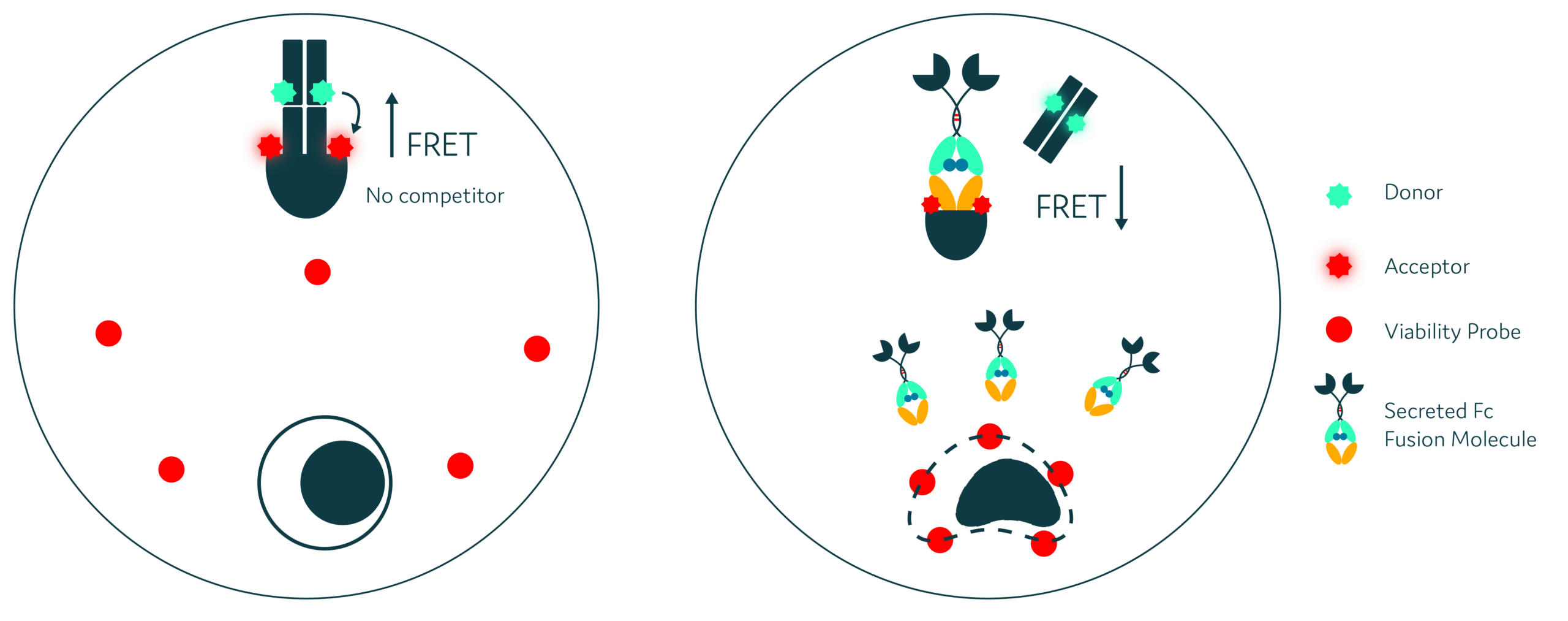
Schematic diagram for Cyto-Cellect® Fc Fusion and Viability Probe assay on Cyto-Mine® Chroma. The FRET donor is labelled with Alexa Fluor 488 and paired with an Alexa Fluor 594-labelled acceptor. Secretion of human Fc fusion molecules reduces the FRET signal as donor is displaced. Binding of the Viability Probe to apoptotic or dying cells provides a simultaneous cell-viability readout.
Have you read enough to want the full protocol?
If yes, simply click the button, and fill in the form to download the complete protocol and start using Cyto-Cellect® Fc Fusion in combination with Viability Probe on Cyto-Mine® Chroma.
Otherwise, keep reading if you need a little more information about the workflow and some of the key benefits first!
How it Works:

Step 1
Prepare Cyto‑Cellect® Fc Fusion assay and viability probe
Step 2
Load sample into Cyto-Cartridge® and start Cyto-Mine® Chroma run:
- Cells are encapsulated into uniform picodroplets with both probe systems
Step 3
Incubation & FRET Signal Generation:
- Measurable emission signal generated
Step 4
Gating & Sorting:
- Gate 1: Exclude non‑viable cells
- Gate 2: Include IgG‑secreting viable cells
Step 5
Dispense viable Fc fusion-secreting cells
Key Benefits:
1) Simultaneous secretion and viability screening
Measure Fc fusion secretion and cell viability in the same picodroplet, allowing high-quality secretors to be selected while dead or dying cells are excluded
2) Direct identification of Fc fusion-secreting cells
The competition FRET assay detects secreted molecules containing the human IgG Fc region through a measurable reduction in FRET signal
3) Improved quality of sorted cells
Excluding Viability Probe-positive cells prevents non-viable secretors from entering the selected population. In the example dataset, approximately 46.9% of the Viability Probe-positive population also displayed a secretion-associated FRET response, illustrating why viability exclusion is important.
4) High-throughput, multiplexed screening
Cyto-Mine® Chroma combines picodroplet encapsulation, incubation, fluorescence detection, sorting and dispensing in an automated single-cell workflow
5) Compatible with downstream cloning or molecular analysis
Selected viable secretors can be dispensed into microtiter plates for clonal expansion
Ready for the full protocol now?
Simply click the button, and fill in the form to download the complete protocol and start combining Cyto-Cellect® Fc Fusion with a Viability Probe to screen for viable antigen-specific cells on Cyto-Mine® Chroma.
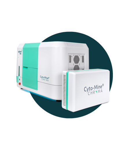
The Next Generation: Cyto-Mine® Chroma
Automate. Accelerate. Analyze millions of cells in a single day.
Think Cyto-Mine®, but supercharged, enabling multiplexing and greater assay flexibility to fit your needs. It means that you can examine vastly greater numbers of cells — and isolate the most valuable ones— with unparalleled precision.