

Protocol: An antigen-specific cell-binding assay for Cyto-Mine® Chroma
Download Now
Protocol Overview
Antigen-specific binding assays are widely used in Antibody Discovery (AbD) workflows to identify antibody-secreting cells (ASC) that produce antibodies capable of recognizing a target antigen. Early identification of antigen-binding clones allows researchers to prioritize target-specific candidates, improving the efficiency of downstream characterization and reducing the time and resources required to identify high-quality antibody leads.
This protocol describes how to design, optimize, and perform an antigen-specific cell binding assay using Cyto‑Mine® Chroma, in which the antigen is expressed on the surface of a target cell.
A simplified schematic of the assay principle is shown in Figure 1. Here both an ASC as well as target cells expressing the antigen of interest, are present in the same picodroplet. As antibodies are secreted, those that recognize the antigen bind to the surface of the target cell within the droplet (Figure 1). Detection is performed using a fluorescently labelled detection antibody that binds to the secreted antibody. In the absence of antigen-specific antibody, the detection antibody will remain unbound to a cell, and the associated fluorescent signal is diffusely distributed throughout the droplet (Figure 1A)
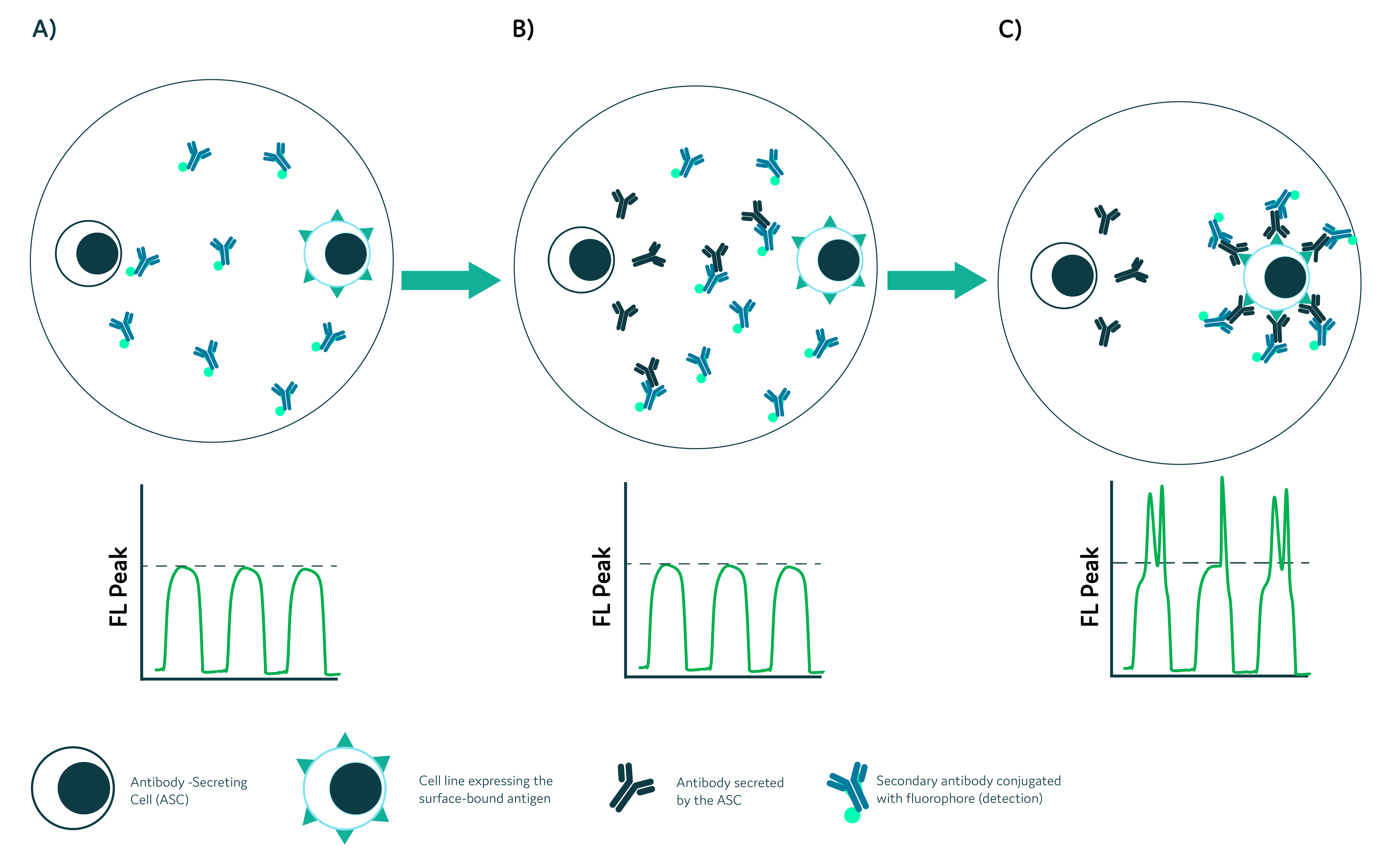
Figure 1. Schematic representation of antigen-specific cell binding assay. A) Antibody-secreting cells are encapsulated together with target cells expressing antigen on the surface, and fluorescently-labeled detection antibody. B) Upon secretion, the primary antibody is recognized by the detection antibody and binds to the antigen on the surface of the cell. C) Binding of the antibody complex on the surface of the target cell results in translocation of some of the fluorescent detection antibody from homogenously dispersed, to localized on the cell surface, which is exhibited as an increase in peak green fluorescence signal. The fluorescence peak data (FL. Peak) is shown for three sequential droplets passing the detection point. The dotted line shows the background peak signal, with peaks appearing above this line indicating binding event.
Following secretion of the antigen-specific antibody in the droplet, the detection antibody binds to the secreted antibody (Figure 1B) and the resulting complex accumulates on the surface of the target cell, generating a localized fluorescence signal (Figure 1C). In Cyto-Mine® Chroma, this signal can be distinguished from the diffuse fluorescence generated by cell – unbound detection antibody distributed throughout the droplet (Figure 1, FL Peak).
This protocol builds on the principles outlined above and takes advantage of the multiplexing capabilities of Cyto-Mine® Chroma to enable highly sensitive detection of antigen-specific ASC (Figure 2). In addition to the use of the fluorescent detection antibody described in Figure 1, both ASC as well as target cells are fluorescently labelled to improve signal discrimination and overall assay performance.
We also provide details of how you can adapt the protocol to exclude non-viable cells, by incorporating our Viability Probe or alternative cell-labeling strategies as appropriate for the experimental system.
Alternative protocols for detecting antibody binding to soluble antigen (FRET assay) or antigen-coated beads are available in our Knowledge Hub.
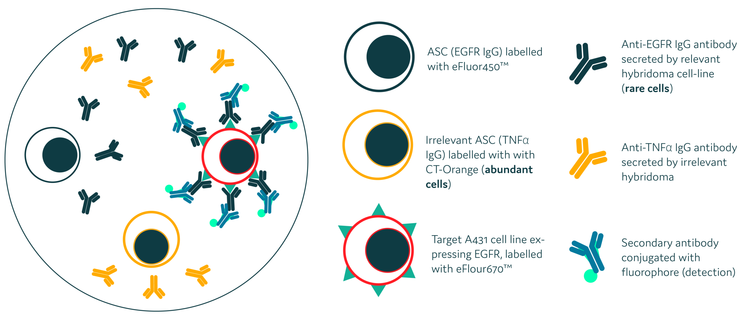
Figure 2. Schematic representation of the antigen-specific assay used in the protocol: Three types of cells were stained and prepared for co-encapsulation in picodroplets: target A431 cells (over-expressing Epidermal growth factor receptor (EGFR)) were stained with eBioscience™ Cell Proliferation Dye, eFluor™670; hybridoma cells, secreting anti-human EGFR antibodies, were stained with eBioscience™ Cell Proliferation Dye eFluor™ 450, and an irrelevant hybridoma cell-line (secreting irrelevant anti-human TNF-α antibodies) was stained with Cell Tracker Orange CMRA. For detection of antigen-specific binding of anti-EGFR Ab to the surface of target cells, anti-mouse AF488-conjugated antibody was added to the reaction.
Have you read enough to want the full protocol?
If yes, simply click the button, and fill in the form to download the complete protocol.
Otherwise, keep reading if you need a little more information about the workflow and some of the key benefits first!
How it Works:

- Prepare cells: Stain the ASC and the target cells
- Prepare sample for Cyto-Mine® Chroma: Prepare mixture of ASC and target cells with anti-mouse AF488-conjugated detection antibody in Encapsulation Medium.
- Load sample into Cyto-Cartridge® and start Cyto-Mine® Chroma run:
a) Cell Encapsulation – ASCs and target cells are encapsulated into uniform picodroplets with fluorescent detection antibody
b) Incubation & Signal Generation – Secreted antibodies bound by the detection antibody are captured on target cells, generating a measurable localized emission signal
c) Gating & Sorting –- Gate 1: Exclude fused droplets
- Gate 2: Include relevant ASC
- Gate 3: Include target cells showing localized antigen-specific binding event
- Dispense antigen-specific ASC for downstream analysis (e.g. RT-PCR)
Key Benefits:
1) Detection of antigen-specific Ab using target cells expressing the native antigen
Using target cells that naturally express the antigen helps preserve its native conformation and membrane context, enabling selection of antibodies that recognise biologically relevant, surface-accessible epitopes
2) Early Identification of Rare Clones
Capture low-frequency antigen-specific ASCs early in the discovery pipeline
3) High Throughput detection of Antigen-specific clones
Screen millions of cells in a single run, reducing development timelines and costs
4) Reduced false positive / background noise
Step-by-step instructions for sample preparation to reduce false positive responses during detection and isolation of clones using Cyto-Mine® Chroma
5) High Specificity
Antigen-specific binding of the secreted antibody in a droplet, results in a clear and localized fluorescence signal detectable on Cyto-Mine® Chroma, supporting detection of highly valuable hits
5) Flexible & Adaptable
The assay may be optimized for use with different target cells and ASC to suit your application
Ready for the full protocol now?
Simply click the button, and fill in the form to download the complete protocol to screen for antigen-specific cells on Cyto-Mine® Chroma.
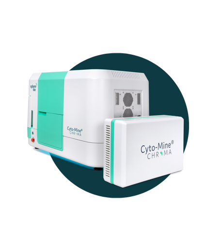
The Next Generation: Cyto-Mine® Chroma
Automate. Accelerate. Analyze millions of cells in a single day.
Think Cyto-Mine®, but supercharged, enabling multiplexing and greater assay flexibility to fit your needs. It means that you can examine vastly greater numbers of cells — and isolate the most valuable ones— with unparalleled precision.