
Protocol: Antigen-Specific FRET Assay for Cyto-Mine® Platforms
Download NowWhy Use This Protocol?
Following this protocol enables high-throughput, efficient, and accurate detection and isolation of rare, antigen-specific antibody-secreting cells early in the antibody discovery workflow. Combining an antigen-specific FRET assay with the Cyto-Mine®’s platform shortens overall discovery timelines and supports earlier identification of the most promising drug candidates.
Key Benefits:
1) Early Identification of Rare Clones
Capture high-value antibody-secreting cells early in the pipeline
2) Accelerated Discovery
Screen millions of cells in a single run, reducing development timelines and costs
3) High Specificity
Directly links antigen-binding function to individual antibody secreting cells, eliminating non-specific selection
4) Reproducible
FRET-based detection delivers clear, measurable signals for confident decision-making
5) Flexible & Adaptable
Easily tailored for different antigens and antibody classes
Protocol Overview
The Antigen-Specific Förster Resonance Energy Transfer (FRET) Assay is a cutting-edge method for detecting antigen-specific antibody secretion at the single-cell level. Designed for use with Cyto-Mine® platforms, this protocol combines fluorescent probe technology, microfluidic encapsulation, and automated sorting to streamline antibody discovery workflows.
In this protocol, TNF-α serves as a model antigen. Hybridoma cells secreting anti-TNF-α antibodies are encapsulated into picodroplets containing fluorescently labelled TNF-α (donor probe) along with fluorescently labelled anti-mouse IgG antibody (acceptor probe). When a secreted antibody binds to TNF-α, the close proximity of the donor-acceptor fluorophores induces FRET, generating a measurable emission signal detected by the Cyto-Mine®’s integrated optics. Positive droplets are automatically sorted for subsequent recovery and characterization (Figure 1).
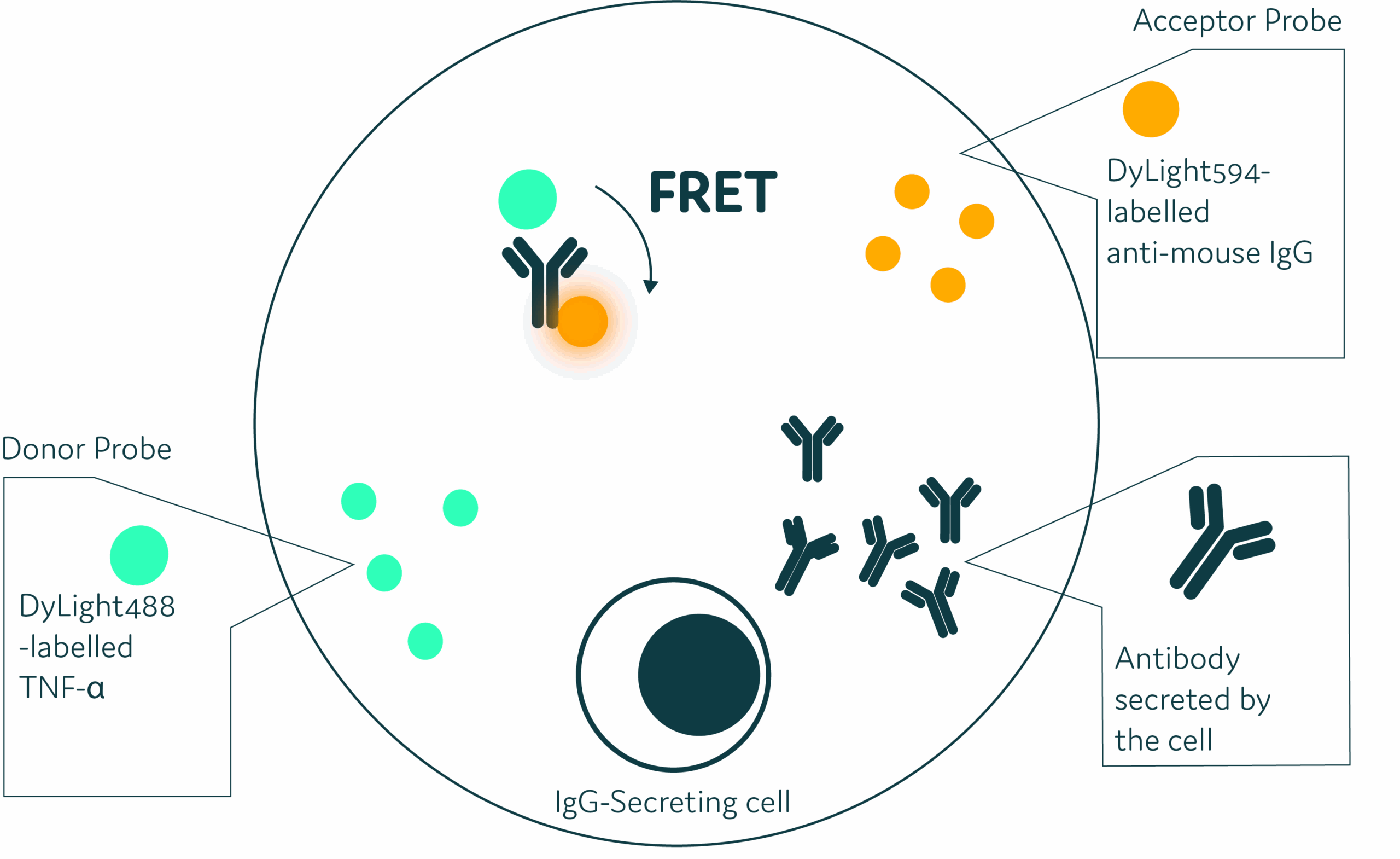
Figure 1: Schematic diagram for antigen-specific specific Förster Resonance Energy Transfer (FRET) assay. This method directly evaluates antigen-specificity of secreted antibody by using FRET, whereby the FRET donor (DyLight488 labelled antigen) is paired with a FRET acceptor (DyLight594 labelled anti-mouse IgG). A FRET response is only detectable when the two FRET probes bind antigen-specific secreted antibody and form a three-molecule complex.
How it Works:
Step 1
Label Antigen with fluorescent dyes
Step 2
Optimize FRET Probe ratios using plate reader analysis
Step 3
Encapsulate single cells in picodroplets with optimized probe ratio
Step 4
Detect and sort positive droplets automatically
Step 5
Dispense antigen-specific ASC for downstream analysis (e.g. RT-PCR)

Want the full protocol?
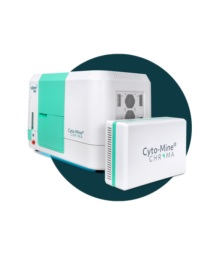
Download NowThe Next Generation: Cyto-Mine® Chroma
Automate. Accelerate. Analyze millions of cells in a single day.
Think Cyto-Mine®, but supercharged, enabling multiplexing and greater assay flexibility to fit your needs. It means that you can examine vastly greater numbers of cells — and isolate the most valuable ones— with unparalleled precision.